ACLS H’s & T’s: Reversible Causes of Cardiac Arrest
This article explains the H’s and T’s in ACLS, the reversible causes of cardiac arrest, and the key clinical clues that may help providers identify and address them during resuscitation.

During cardiac arrest, high-quality CPR, timely defibrillation when indicated, and guideline-directed ACLS care remain the immediate priorities. But the resuscitation team must also ask another critical question:
What caused the cardiac arrest — and is the cause potentially reversible?
The H’s and T’s provide a structured framework for identifying conditions that may have contributed to cardiac arrest and may require targeted treatment during resuscitation. The 2025 American Heart Association Adult Cardiac Arrest Algorithm specifically includes treatment of reversible causes as part of ongoing cardiac arrest management.
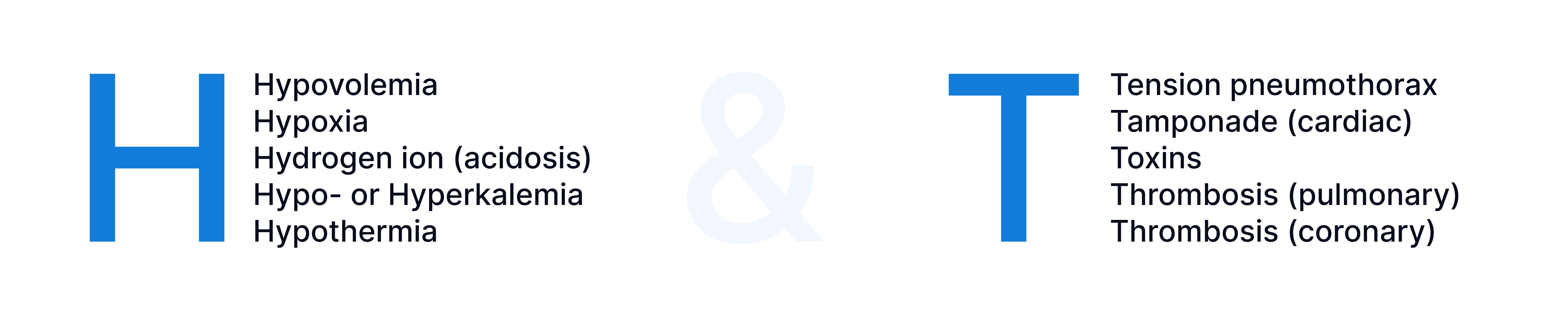
What Are the H’s and T’s in ACLS?
The H’s
Hypovolemia
Hypoxia
Hydrogen ion (acidosis)
Hypokalemia or Hyperkalemia
Hypothermia
The T’s
Tension pneumothorax
Cardiac tamponade
Toxins
Pulmonary thrombosis
Coronary thrombosis
These causes should not be treated as a checklist that automatically triggers an intervention. Instead, providers use the patient’s history, clinical circumstances, examination findings, rhythm, and available diagnostics to determine which causes are plausible and which may require immediate action.
For pediatric resuscitation, the PALS reversible causes follow a similar framework, with hypoglycemia included as an additional reversible cause. You can learn more in our article on the H’s and T’s in PALS.
Why Reversible Causes Matter During ACLS
An arrhythmia may be the immediate mechanism of cardiac arrest without being the underlying problem.
For example, pulseless electrical activity may occur because effective circulation is being prevented by severe volume loss, pulmonary embolism, tension pneumothorax, or another underlying condition.
Identifying and treating a likely reversible cause can therefore become an important part of resuscitation while the team continues standard ACLS care.
The key practical question is:
Which reversible cause best fits this patient, and is there an intervention that could change the course of the arrest?
Using the Secondary Assessment to Look for a Cause
The search for reversible causes can begin as soon as information can be gathered without compromising CPR, defibrillation, airway management, or other critical interventions.
Useful information may come from:
The history. Consider what happened immediately before the arrest, recent symptoms, known medical conditions, medications, dialysis history, procedures, trauma, possible toxic exposure, or significant fluid or blood loss.
The physical examination. Findings such as severe bleeding, signs of trauma, abnormal breath sounds, or other clues may help narrow the differential diagnosis.
Available monitoring and diagnostics. Rhythm information, capnography, laboratory values, and selected imaging can sometimes contribute useful information.
Point-of-care ultrasound may also be considered by experienced professionals when it can be performed without interrupting CPR. However, the AHA notes that its usefulness for diagnosing reversible causes during cardiac arrest is not well established, so ultrasound findings should be interpreted within the broader clinical context.
The H’s
1. Hypovolemia
Hypovolemia refers to an inadequate circulating blood volume. Severe hemorrhage or substantial fluid loss can reduce venous return and cardiac output to the point that effective circulation cannot be maintained.
Consider hypovolemia when the history or circumstances suggest:
Major hemorrhage
Significant trauma
Severe dehydration or fluid loss
Recent bleeding or surgery
Other conditions associated with major volume depletion
During resuscitation, treatment is directed toward the underlying source of volume loss. Depending on the cause and clinical setting, this may include hemorrhage control and appropriate volume or blood-product replacement according to established protocols.
The important point is not simply to recognize “low volume,” but to determine why the patient has lost effective circulating volume and whether that cause can be corrected.
2. Hypoxia
Hypoxia occurs when inadequate oxygen reaches the tissues and can be both a cause and consequence of cardiac arrest.
Potential causes include airway obstruction, respiratory failure, inadequate ventilation, misplaced airway equipment, or interruption of oxygen delivery.
During ACLS, assess:
Is the airway open?
Is ventilation producing appropriate chest rise?
Is oxygen being delivered effectively?
If an advanced airway is present, is its position confirmed?
Is an equipment problem interfering with ventilation?
During adult cardiac arrest, the AHA states that maximal feasible inspired oxygen may reasonably be used when supplemental oxygen is available. If an advanced airway is placed, continuous waveform capnography is recommended to confirm and monitor endotracheal tube placement. Airway procedures should not create unnecessary interruptions in chest compressions.
3. Hydrogen Ion Excess (Acidosis)
Significant acidosis can accompany prolonged cardiac arrest, severe shock, renal dysfunction, sepsis, and other major metabolic disturbances.
During cardiac arrest, acidosis is often related to inadequate circulation and ventilation. Improving high-quality CPR, oxygenation, ventilation, and perfusion therefore remains fundamental.
One important point for ACLS providers is that routine sodium bicarbonate administration during adult cardiac arrest is not recommended. The 2025 AHA guidelines maintain this recommendation because routine administration has not demonstrated improved outcomes. Bicarbonate may still have a role in selected special circumstances addressed separately in the guidelines.
So, “acidosis” on the H’s and T’s list should not be interpreted as an automatic indication to administer bicarbonate.
4. Hypokalemia or Hyperkalemia
Abnormal potassium levels can produce severe disturbances in cardiac electrical activity and may contribute to cardiac arrest.
Possible clues include:
Renal failure
Dialysis
Medications that alter potassium levels
Potassium supplementation
Crush injury
Previous laboratory abnormalities
ECG changes compatible with an electrolyte disturbance
Hyperkalemia deserves particular caution. The 2025 AHA Special Circumstances guidelines state that the effectiveness of IV calcium, sodium bicarbonate, and insulin with glucose during cardiac arrest caused by suspected hyperkalemia is not well established. Inhaled β2-agonists are not recommended during hyperkalemic cardiac arrest.
This does not mean that hyperkalemia should be ignored. It means treatment should be based on the clinical situation, available evidence, and the applicable resuscitation protocol rather than assuming that a single medication is universally effective.
5. Hypothermia
Severe environmental hypothermia can itself cause cardiac arrest.
Unlike many other arrest situations, prolonged resuscitation may still result in good neurological recovery in selected patients with severe hypothermia.
The AHA recommends that adults with life-threatening environmental hypothermia and no obvious lethal injury receive full resuscitative measures concurrently with rewarming. Extracorporeal life support may be reasonable for rewarming adults in hypothermic cardiac arrest when available.
Possible clues include:
Significant cold exposure
Very low core temperature
Wet clothing or prolonged environmental exposure
Cardiac arrest occurring in a cold environment
Hypothermic cardiac arrest also has specific considerations regarding defibrillation, epinephrine, transport, and extracorporeal rewarming, so providers should follow dedicated hypothermia guidance rather than treating it exactly like a normothermic arrest.
The T’s
1. Tension Pneumothorax
A tension pneumothorax can obstruct venous return to the heart and cause rapid cardiovascular collapse.
Clinical circumstances that may increase suspicion include:
Significant chest trauma
Recent thoracic procedures
Sudden difficulty ventilating the patient
Markedly asymmetric breath sounds
Other findings compatible with obstructive shock
Because tension pneumothorax can be rapidly fatal yet potentially reversible, it should remain in the differential diagnosis when the clinical circumstances fit.
Management should follow emergency and trauma protocols for suspected tension pneumothorax while minimizing interruption of CPR.
2. Cardiac Tamponade
Cardiac tamponade occurs when fluid or blood within the pericardial space creates enough pressure to impair cardiac filling.
Possible contexts include:
Penetrating chest trauma
Recent cardiac procedures
Known pericardial effusion
Other circumstances associated with pericardial bleeding
POCUS may sometimes help identify findings associated with tamponade, but its use during cardiac arrest should not prolong pauses in chest compressions, and the AHA emphasizes that the diagnostic value of POCUS for reversible causes remains imperfect.
When tamponade is strongly suspected, definitive management depends on the clinical context and available expertise.
3. Toxins
Poisoning and drug exposure can produce cardiac arrest through many different mechanisms.
Possible clues include:
Known or suspected overdose
Medication errors
Information from family, EMS, or witnesses
Medication containers or environmental exposures
Clinical findings consistent with a particular poisoning
There is no single “toxin treatment” within ACLS. Management depends on the substance involved.
The 2025 AHA Special Circumstances guidelines provide toxin-specific recommendations for multiple exposures, including opioids, β-blockers, calcium channel blockers, digoxin, cyanide, sodium-channel blockers, local anesthetic toxicity, sympathomimetics, and others.
When a toxin is suspected, the team should identify the likely exposure as quickly as possible and use appropriate toxin-specific treatment alongside standard resuscitation.
4. Pulmonary Thrombosis
Pulmonary thrombosis generally refers to a massive pulmonary embolism severe enough to cause circulatory collapse or cardiac arrest.
A large pulmonary embolism can dramatically increase pulmonary vascular resistance, impair right ventricular function, and ultimately prevent adequate circulation.
Possible clues include:
Sudden unexplained collapse
Known or previous pulmonary embolism
Venous thromboembolism risk factors
Severe preceding respiratory or hemodynamic deterioration
Evidence suggesting right ventricular strain
For adults with confirmed PE as the cause of cardiac arrest, the AHA states that systemic fibrinolysis, surgical embolectomy, and percutaneous mechanical embolectomy are reasonable treatment options. ECLS is also reasonable for cardiac arrest caused by confirmed or suspected PE. When PE is suspected but not confirmed, systemic fibrinolysis may be considered.
The appropriate strategy depends heavily on the individual patient and the resources available.
5. Coronary Thrombosis
Acute coronary occlusion can cause myocardial ischemia, ventricular arrhythmias, and cardiac arrest.
Before arrest, clues may include symptoms or findings suggestive of acute coronary syndrome. After ROSC, the ECG and the patient’s hemodynamic status become particularly important.
The 2025 AHA post-cardiac arrest guidelines recommend emergent coronary angiography after cardiac arrest when a cardiac cause is suspected and persistent ST-segment elevation is present.
Emergency angiography can also be reasonable in selected patients without ST elevation when there is cardiogenic shock, recurrent ventricular arrhythmia, or evidence of significant ongoing myocardial ischemia. Routine immediate angiography is not recommended for otherwise stable comatose patients without these features.
A Quick H’s and T’s Reference
Reversible Cause | Examples of Clues | General Treatment Direction |
|---|---|---|
Hypovolemia | Hemorrhage, trauma, major fluid loss | Identify and correct volume loss; control bleeding when present |
Hypoxia | Airway or ventilation problem, respiratory failure | Restore effective oxygenation and ventilation |
Hydrogen ion / acidosis | Prolonged arrest, shock, severe metabolic illness | Optimize CPR, ventilation and perfusion; address underlying cause |
Hypo-/hyperkalemia | Renal failure, dialysis, medications, abnormal ECG/labs | Identify and manage the electrolyte disorder according to applicable guidance |
Hypothermia | Cold exposure, low core temperature | Continue resuscitation while rewarming; consider specialized hypothermia pathways |
Tension pneumothorax | Trauma, thoracic procedure, ventilation difficulty | Treat suspected tension physiology promptly according to emergency protocols |
Cardiac tamponade | Trauma, cardiac procedure, pericardial disease | Identify and address obstructive pericardial pressure when indicated |
Toxins | Overdose, exposure, medication history | Use toxin-specific resuscitation and antidotal guidance |
Pulmonary thrombosis | PE risk, sudden collapse, obstructive physiology | Consider PE-specific therapies when confirmed or strongly suspected |
Coronary thrombosis | ACS presentation, ischemia, post-ROSC ECG changes | Follow post-cardiac-arrest and coronary reperfusion pathways |
How Should the H’s and T’s Be Used During ACLS?
The value of the H’s and T’s is not simply memorizing ten terms. Their purpose is to provide the resuscitation team with a shared diagnostic framework.
While standard ACLS interventions continue, the team can repeatedly ask:
What happened immediately before the arrest?
Is there evidence of respiratory failure or major blood loss?
Does the patient’s medical history suggest an electrolyte disturbance?
Was there trauma or a recent procedure?
Could this be an overdose?
Could an obstructive cause such as PE, tamponade, or tension pneumothorax explain the arrest?
Is there evidence suggesting an acute coronary cause?
The most useful reversible cause is not necessarily the first one on the list. It is the one that best fits the patient’s clinical situation and offers an actionable intervention.
Key Takeaway
The H’s and T’s help ACLS providers systematically consider potentially reversible causes of cardiac arrest. They should support (not distract from) high-quality CPR, appropriate defibrillation, airway and medication management, and the other core components of ACLS.
The foundation of adult cardiac arrest resuscitation remains high-quality CPR, early defibrillation when indicated, guideline-directed pharmacologic treatment, and appropriate airway management. Investigation of reversible causes should occur alongside these priorities rather than delaying them.
Was this article helpful?
Quick tip: Let us know if this article was helpful and discover a special discount code for your upcoming certification.
Preparing for ACLS Certification?
Get officially certified in ACLS online with a streamlined, flexible course designed for busy healthcare professionals. Complete it in under an hour and receive your provider card instantly.
Get Certified